Learn More
iTrace™: Elevating Small Aperture IOL Success
iTrace™ bridges patient selection, surgical precision, and post-op validation—before and after Small Aperture IOL implantation. Its proprietary ray tracing technology gives you a high-resolution view of visual quality, helping your team optimize outcomes.

iTrace Separates Cornea from Crystalline Lens
Evaluate visual quality from the cornea independently of the crystalline lens
Understand how the cornea may affect the patients vision post-operatively
Let’s Review: What is Ray Tracing?
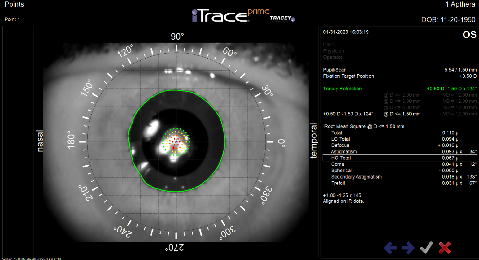
Sends up to 256 individual and sequential laser beams through a 1–8 mm pupil in under ¼ second
Each beam is traced to the macula to map its path and impact.
Ray tracing combined with corneal topography data isolates how the cornea and crystalline lens affect each beam
Automatically calculates refraction and higher-order aberrations for precise analysis
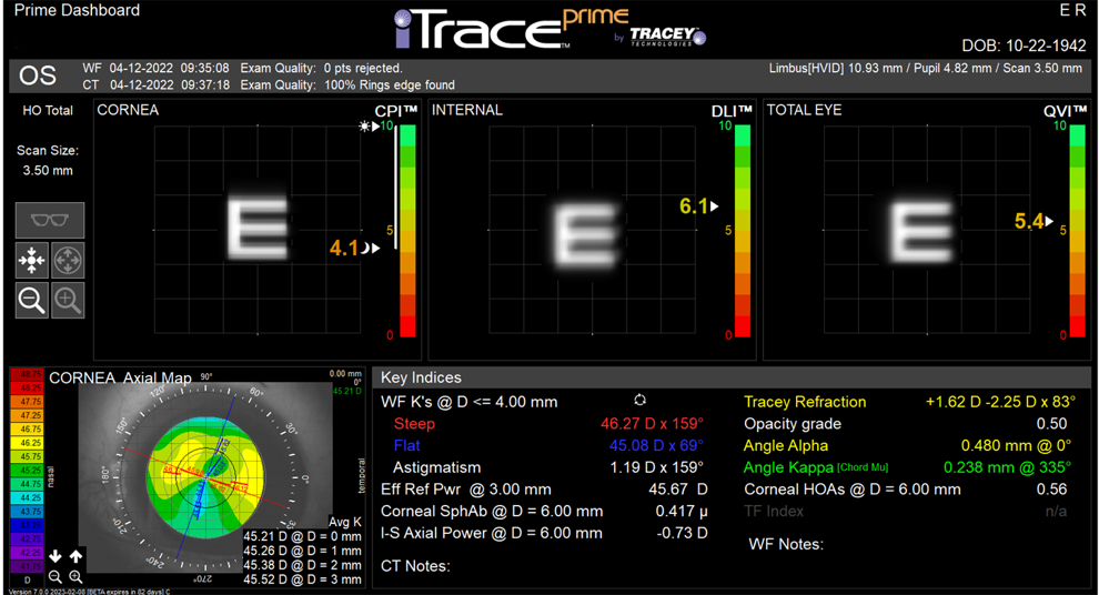
CPI, DLI, QVI, and TFI
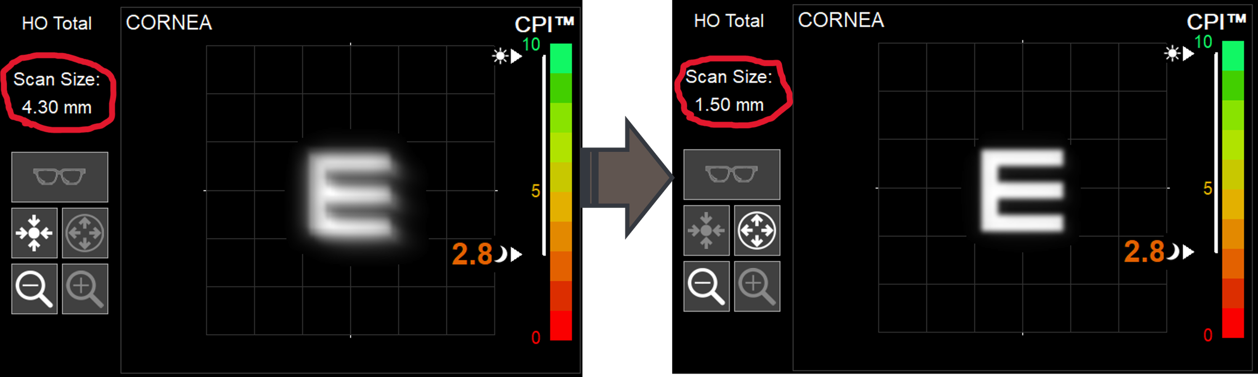
Corneal Performance Index (CPI)

The Letter “E” in the display reflects Quality of Vision from the cornea
The CPI is a 0 to 10 metric to screen for premium lens candidacy: poor corneal performance can indicate that a light filtering small-aperture IOL may be beneficial for the patient.
Dysfunctional Lens Index (DLI)
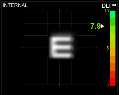
The Letter “E” in the display reflects Quality of Vision from the crystalline lens
The DLI is a 0 to 10 objective metric that can indicate the soonest and best time to exchange the lens, either as RLE or Cataract Surgery.
Quality of Vision Index (QVI)
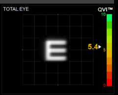
The Letter “E” simulates how a patient sees normally without correction at the largest pupil size measured.
The QVI is a 0 to 10 metric that objectively quantifies the quality of the patient’s overall vision based on the whole eye’s aberration profile.
Tear Film Index (TFI)
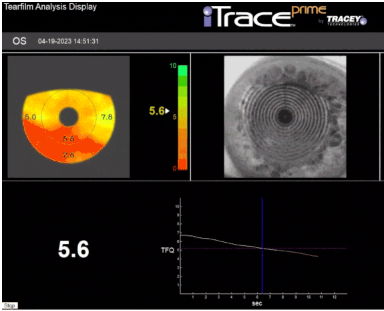
The TFI is a 0 to 10 metric that objectively quantifies the quality of the tear film function.
Know if tear film function will provide clear vision or if it needs to be managed first
Pre-Op Simulation of Small Aperture Vision
Educate Patients – Simulate how a small aperture optic filters unfocused light from the cornea
Unlock Post-Op Clarity - Small Aperture Autorefraction
Only device to auto-refract through a small aperture
Assess lens centration and target accuracy
Diagnose refractive surprises
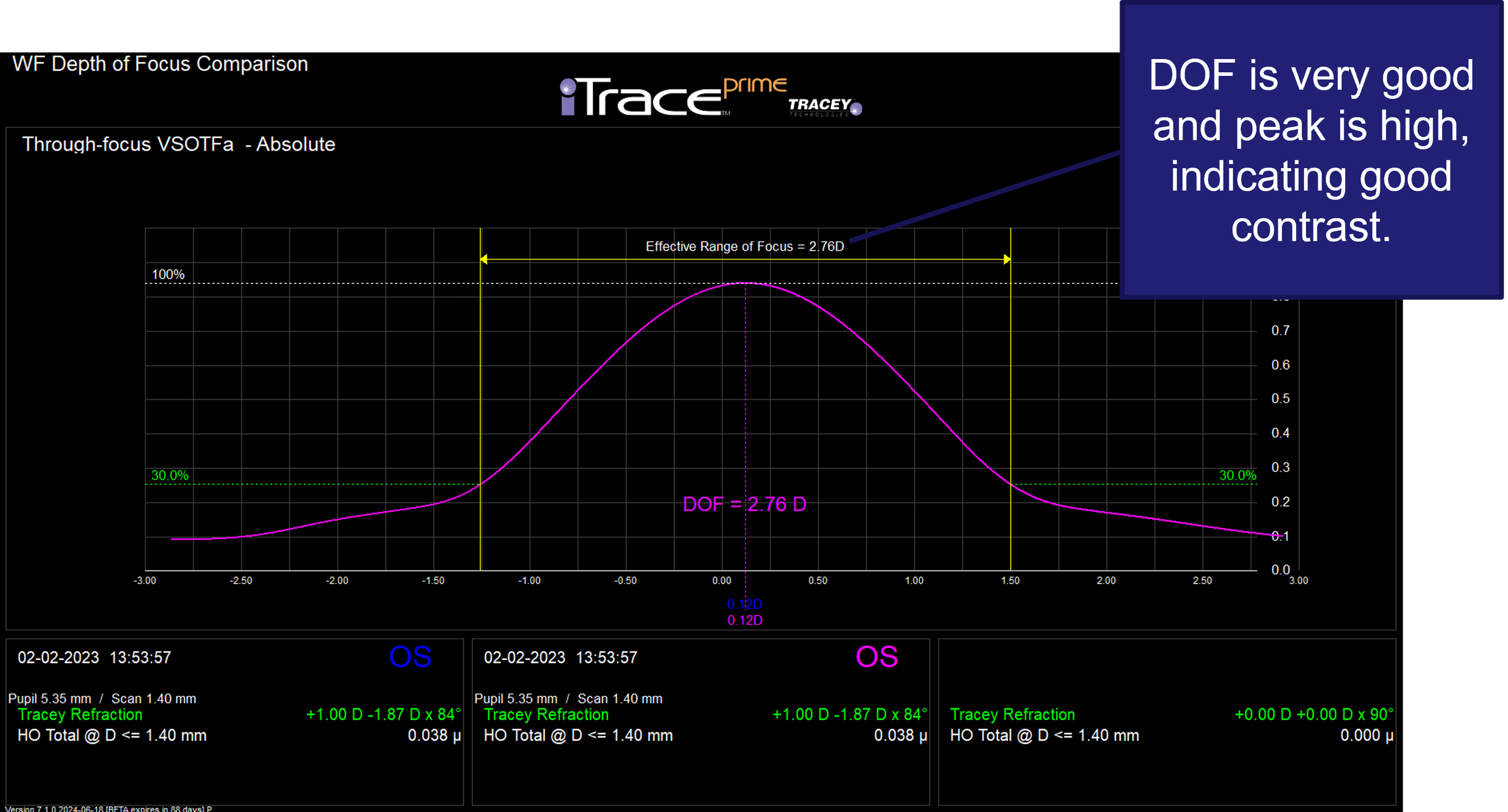
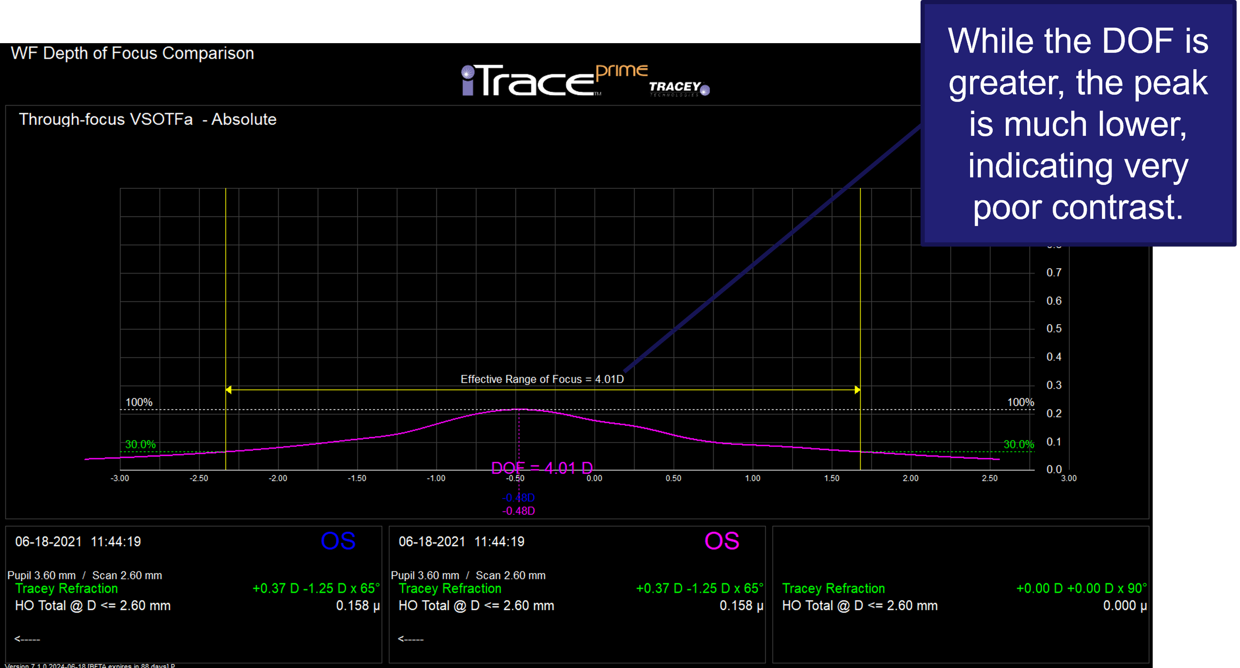
Visualize the full depth-of-focus curve
Post-op changes in QVI can signal PCO Formation.
Defocus Curve Analysis Post-OP
Small Aperture IOL
Trifocal IOL